- Synonyms
- Hematopoietic Growth Factor, MCGF, Multipotential Colony-stimulating Factor, P-cell-stimulating Factor
- Source
- Escherichia coli.
- Molecular Weight
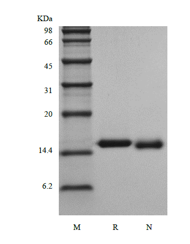
- Approximately 14.0 kDa, a single non-glycosylated polypeptide chain containing 124 amino acids.
- AA Sequence
- APMTQTTSLK TSWAKCSNMI DEIITHLNQP PLPSPDFNNL NEEDQTILVE KNLRRSNLEA FSKAVKSLQN ASAIESILKN LPPCLPMATA APTRPPIRIT NGDRNDFRRK LKFYLKTLEN EQAQ
- Purity
- > 98 % by SDS-PAGE and HPLC analyses.
- Biological Activity
- Assay #1: Fully biologically active when compared to standard. The ED50 as determined by the dose-dependent stimulation of the proliferation of murine NFS-60 cells is less than 5.0 ng/ml, corresponding to a specific activity of > 2 × 10⁵ IU/mg.
Assay #2: The ED50 as determined by a cell proliferation assay using human TF-1 cells is less than 0.05 ng/ml, corresponding to a specific activity of > 2.0 × 10⁷ IU/mg.
- Physical Appearance
- Sterile Filtered White lyophilized (freeze-dried) powder.
- Formulation
- Lyophilized from a 0.2 µm filtered concentrated solution in PBS, pH 7.4, 5 % trehalose.
- Endotoxin
- Less than 1 EU/µg of rRhIL-3 as determined by LAL method.
- Reconstitution
- We recommend that this vial be briefly centrifuged prior to opening to bring the contents to the bottom. Reconstitute in sterile distilled water or aqueous buffer containing 0.1 % BSA to a concentration of 0.1-1.0 mg/ml. Stock solutions should be apportioned into working aliquots and stored at≤ -20 °C. Further dilutions should be made in appropriate buffered solutions.
- Stability & Storage
- Use a manual defrost freezer and avoid repeated freeze-thaw cycles.
- 12 months from date of receipt, -20 to -70 °C as supplied.
- 1 month, 2 to 8 °C under sterile conditions after reconstitution.
- 3 months, -20 to -70 °C under sterile conditions after reconstitution.
- Usage
- This material is offered by Shanghai PrimeGene Bio-Tech for research, laboratory or further evaluation purposes. NOT FOR HUMAN USE.
- SDS-PAGE
- Reference
- 1. Yang YC, Ciarletta AB, Temple PA, et al. 1986. Cell. 47:3-10.
2. Otsuka T, Miyajima A, Brown N, et al. 1988. J Immunol. 140:2288-95.
3. Dorssers L, Burger H, Bot F, et al. 1987. Gene. 55:115-24.
4. Feng Y, Klein BK, McWherter CA. 1996. J Mol Biol. 259:524-41.
- Background
- Interleukin-3 (IL-3) is an interleukin, a type of biological signal (cytokine) which is encoded by the IL-3 gene located on chromosome 5 and produced primarily by activated T cells beside human thymic epithelial cells, activated murine mast cells, murine keratinocytes and neurons/astrocytes. The protein acts in hematopoiesis by controlling the production, differentiation, and function of 2 related white cell populations of the blood, the granulocytes and the monocytes-macrophages. The rhesus macaque IL-3 reported to be a monomer, as it is known, contains 124 amino acids residues which is a single non-glycosylated polypeptide. Specifically, recombinant rhesus macaque, human and murine IL-3 share low homology.


 COA Application


 Home
Home Product Datasheet
 MSDS
MSDS
 Screening Products